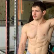
Alessandro Mainente Posted May 24, 2012 Share Posted May 24, 2012 some days ago i've followe a seminar (wich was the result of an extensive research and study) in my university about the need to use the functional training for the rehabilitation process also in person who suffer of obesity or need to be stronger for future strength elementsthe seminar covered only one case of the application of funtional training. btw i had the opportunity to ask to the guy who did the relation about the instruments that were used in this study, he was talking about trx, medica ball and of course gymnatics rings and something more (bar, proprioception tables etc)he wasn't specific about the exercises but he talked about plank and L-sit progression for some people.The seminar was offered also in papers so i have copied the biggest and relevant part of the study here, i hope you can enjoy it! INTRODUCTIONIn many popular and professional publications, theterm functional training, 3D training or spatial exerciseis often encountered. Functional training from a clinicalanatomicalperspective and its application in clinicalpractice is dependent on a sound knowledge of clinicalanatomy. Functional training highlights a need for furtheringanatomical knowledge in the postgraduate educationof physicians from various fields of clinical practice, e.g.in sports medicine and physiotherapy.Functional training is a suitable alternative to powerexercise and stems from rehabilitation and physiotherapyroutines. It is modelled on the basis of the patient’s functionalimpairment and muscle function test results. Theaim of 3D training is not only to improve physical fitnessand muscular coordination, it is also recommended forall age groups in healthy as well as indisposed individuals.Many of its exercises are based on common everydayactivities which require coordination and fluid movementto a greater or lesser extent, such as walking, running,lifting an object, etc. During such daily activities we callinto action many muscle groups, with increased demandson neuromotor coordination and maintenance of bodyposture.We have been using 3D training in our Pediatric Clinicfor the therapy of obese children. We believe it has widerapplication in various clinical fields. However, search ofthe traditional medical data bases reveals a dearth of information.This review aims to provide elementary informationon the principles, roles, methods and applicabilityof 3D training.METHODSDatabase search using the lemmatization methodand data bases: Web of Knowledge, Web of Science,UpToDate, PubMed and Google Scholar for the years2005-2011. Given the dearth of information in traditionalmedical databases, these were available options.We examined studies that focused on the possibilitiesof using functional 3D training in clinical practice basedon implementing the anatomical findings. As this is afairly new method, so far not used as standard in generalclinical practice, we were only able to carry out analysisof a limited number of studies. Our experiences are presentedin a case report which correlates with availablereferences.RESULTSFunctional trainingFunctional training is a synergetic involvement ofseveral muscle groups simuůtaneously enabling trainingthat is more productive. The most commonly used toolsfor 3D training are: free pulleys, single-handed weights, amedicine ball, a large ball, exercise bands, imbalance pads,bags with sand or water, GRAVITY program in rehabilitationon Power Tower system and the very effective armynavy system TRX- Training Resistance Exercise..A prerequisite for this training is quality of perceivedmovement rather than isolated muscle function. Duringactivities of daily living, the body does not move in isolatedpatterns. It moves as an integrated unit, be it functionalor dysfunctional It strives to create such individualstructure of exercises that help carry out common dailyactivities with greater efficiency and lower risk of injury.At the same time, the training leads to greater muscularbalance and strengthening of the spine stabilizationsystem; it has positive impact on articular stability andsignificantly contributes to preventing musculoskeletalinjuries. Functional training restores and enhances theway the body performs everyday physical activities andmemorizes such muscular functions.All these muscles create only a single subsystem ofthe deep stabilization system, known as active. The passiveelement is formed by the vertebrae, ligaments andintervertebral discs. The stability of the spine is impactedby the neural element via the afferent supply from receptors,and subsequent control of the active element.Imbalance of one of their elements can cause a) immediatecompensation – function normalisation, b) long-termadaptation process of one or several subsystems – withfunction normalisation, but with a change in the stabilizationsystem, c) disabling of one or several elementsof some system – with total imbalance, which leads toe.g. the painful syndrome of low back pain (LBP) – inthese patients we find variations in stabilization of musclefunction in comparison with the development model ofstabilization. In patients with LBP, greater damage inthe thoracic area of Musculus erector spinae (MES) incomparison to the lumbar area of MES was proven withan EMG examination. Targeted interference of the deepstabilization system in chronic vertebrogenic difficultiesis therefore the main therapeutic procedure and enablestargeted focus on the damaged area. One caveat is thatopinions on the character of these exercises differ.The therapeutic methods can be used not only as partof physiotherapy, for the therapy of already developedmuscular and neuromuscular imbalance, but also as preventionin reconditioning and training of athletes, especiallyin sports with significantly asymmetric degree ofload in the lumbar spine area. This fact was confirmed bythe Renkawitz’s study, on tennis players and proved thedevelopment of neuromuscular imbalance at the L2 andL4 level in connection with asymmetric load, exhibitedby altered electric activity of MES measured by means ofEMG examination.The National Academy of Sports Medicine (NASM),which was founded in 1987 in Chicago, specifies functionaltraining as a modern method of exercise involvingcoordinated precision movement in the stages of acceleration,deceleration and stabilization of the postural musclegroups in all three axes of the Cartesian coordinate system13.Schofftal (2010) found a significantly greater muscleactivity examined with electromyographic (EMG) of theinternal obliques (IOs), during a prone V-up on TRX comparedto various abdominal isometric exercises.Since physical training can be an important methodfor preventing falls e.g. in patients with neurodegenerativedisease, and given the positive effects of exercise onhealthy articular cartilage it can be protective against theonset of osteoarthrosis.Functional training performed on unstable surfacesmight best be utilized during after injury rehabilitation orduring in-season training to maintain core endurance levelsand reduce the incidence of injuries. Fitzgerald, Axe,and Snyder-Mackler found that performance of wobbleboard exercises improved rehabilitation outcomes in subjectswith anterior cruciate ligament (ACL) rupture.During the exercise, we usually work with the weightof our own body, many muscle groups are connected inone fluid exercise movement and we check the correctposture of body’s centre of gravity (COG), flexibility andcoordination of muscle groups, and especially the postureand operation of body’s deep stabilization system, i.e. posturalmuscles during training.The main quantitative physical forces affecting thehuman body are of three types: gravity, the strength ofmuscles and the “third factorâ€, which is known as theforce of physiological impact and deformation forces. Theeffect of these forces on a specific body element is concentratedon a single point, the body’s centre of gravity.The position of such centre of gravity is what absolutelydetermines the stability of a body and enables us tostudy the course of movement and conditions of bodyposture. Change in the body posture or manifestationsof instability are, according to the law of action and reaction,balanced by tonic correction of muscles, wherebyupon incorrect involvement of compensation muscles orupon muscle function impairment, we must renew correctstability of the body through exercise and therebyretroactively effect the body’s centre of gravity.The body’s centre of gravity has no permanent placeas it oscillates according to the movement of its parts.The centre of gravity in basic anatomical position of restis in the median line at L3-L4 in women and L2-L3 inmen, 4.5-5.5 cm ventrally from facies anterior corporisvertebrae.If the body’s centre of gravity is the foundation ofbody’s stability, such stability is then increased by posture,muscle strength and increased mass of the body erectors.One of the most significant ones, activated to the maximumextent during functional training, is musculus erectorspinae (MES).MES is the most extensive and voluminous of the deepstabilization system muscles, the main function of whichis erecting the torso. It belongs to postural muscles thatcontrol and impact active movements of the spine, apartfrom anteflexion due to the antagonist activity of the abdominalmuscles.As for its function, MES is responsible for maintainingstability of the spine and as bilateral action, for erectingthe spine and backward bending of the head. As unilateralaction, it helps in bending of the spine and rotation to theside of an acting muscle. It participates practically in allfundamental and critical movements required for movementof an individual.Deep dorsal muscles belong to a group of indigenousmuscles and they can be divided according to the layersin a dorsal-ventral direction into.MES is in intense and constant interaction with alllayers of the dorsal muscles and impacts the function andefficiency of the abdominal muscles and participates inthem. The system of work and stabilisation of abdominalmuscles was demonstrated by Kapanji on a model of twocircles with equal diameter and on the principle of a rotatinghyperboloid, the surface of which is sunken likea hyperbolic curve. Maikala also describes significantrelations between movements of the spine and vascularsupply to intervertebral discs and muscles of the spine,including MES . The intervertebral disc (IVD)has important mechanical functions such as spine loaddistribution and maintenance of flexibility. IVD degenerationrepresents a major pathological process in low backpain.Correct function of MES plays a significant role inproper function of the entire deep stabilization systemof the spine. It participates in spine stabilization duringstatic load while seated or standing and also during dynamicload when the extremities are in motion; it allowstransfer of forces and load from the area of upper andlower extremities, the pelvis and the upper torso.The muscle stabilization system in the lumbar spinearea comprises the major stabilizers latissimus dorsi m.,gluteus maximus m., erector spinae m., biceps femoris m.,exteral and internal oblique abdominal m, rectus abdominism., which secure direct segmental stability togetherwith the pelvic floor muscles and the diaphragm.These muscles are demonstrated in figures which are sequences of the one 3D exercise. Figures showanatomical muscle teamwork during phases of the movement.CORE trainingThe term “COREâ€, i.e. the centre, in specialised terminologymeans muscles of the trunk in the area of the thoracicand cervical spine, in the lumbar area, area of pelvisand hips – the so called LPHC complex (lumbar-pelvichipcomplex). Any fast and sudden changes in the directionand position of the body are perceived as a changein balance. These approximately 26 muscles stabilize andprovide control over all movements. In special athletictraining the primary target is strengthening the “centreof the bodyâ€. In this area, all movements are initiatedand the body’s centre of gravity – COG – is located here.Such athletic training leads to strengthening of the deepmuscle system, i.e. muscles that are not often involved incommon exercise. Core training also helps in preventinginjuries during athletic as well as everyday movement,backaches, and it improves body posture.Core training is used by professional fitness trainersto render speed and strength abilities, which are reflectedin repeated accelerations, more effective. A firm trunkshould definitely not be omitted, as it plays a great rolein preventing injuries and it is also beneficial in one’spersonal life. These exercises are focused on static musclestrengthening and compensations of muscle imbalances,mainly in the area of trunk, where the entire body is stabilizedand remains in a balanced position for a periodof 15 s/30 s/45 s/60 s.As a result, the deep stabilization muscles are strengthened.In this way we also focus on strengthening phasicmuscles, which have the tendency to slacken and westretch postural muscles that tend to shorten.Training Resistance Exercise (TRX) and Gymnastics RingsThe TRX system and the gymnastic rings are a unique suspension systems, whichuses movement in all three planes for exercising with thepossibility of adjusting the level of difficulty and the incorporationof additional balancing tools. Suspensiontraining is characterized by one or more hands or feetsupported by a single anchor point while the opposite endof the body is in contact with the ground. It uses your ownbody-weight as resistance and takes advantage of stabilityto load and unload exercises. Resistance exercises performedin unstable body positions have been hypothesizedto increase the muscular strength and muscular enduranceof the core musculature, which may translate to morepowerful and efficient movement patterns and less risk.The entire system has great use, apart from in cardioand strength fitness training, mainly in smooth physiotherapyfor muscle imbalance, disorders of knee stability,chronic “low back pain†conditions, strengthening thedeep stabilization system of the body, strengthening thepelvic floor muscles and so on.The results of the three-month suspension trainingstudy of Yang on the diving athletes’ shows, that balancingand core stability capacity were greatly improved, particularlyin the non-stable state, which means the coordinationof their internal muscle and the coordination betweenmuscles were greatly improved. At the same time, bytesting the front and back vertical Jump, it is noted thatthe athletes’ explosive force of lower limbs significantlyimproved. All the improvement in these abilities mayhave a direct impact on improving their special technicalcapabilities. The study showed that physical trainingbased mainly on the suspension method is a new way ofstrength training and it is especially important for theskill-led events such as diving .CASE HISTORYA boy (L.B., 16 years of age) was examined at ourpaediatric obesitology outpatient department.Subjectively: the patient complained of poor physicalfitness, shortness of breath even during moderate exercise(longer walks over a straight terrain, walking up the stairs,etc.). He jad excessive perspiration frequent pains in bothknee joints when idle (VAS3-4) and following physicalactivity (VAS 7).Objective findings: obesitas magna, fine pink stretchmarks on the abdomen and arms. Remaining somaticfindings were physiological.Anthropometric parameters: Height 185 cm, weight:104 kg, BMI: 30.4, waist 99 cm, hips 114 cm, Waist/hipratio: 0.86, arm circumference: 33 cm, thigh circumference66cm, chest circumference at max. inspiration: 110cm, chest circumference at max expiration: 105cm, (differencebetween chest circumference inspiration/expiration:5cm), chest circumference at middle position: 108 cm.Locomotor apparatus examination: Relaxed posturewhen standing, head protuberant forward, shortenedscalene muscles, shortened elevator scapulae musclemore significantly l.dx, shoulders position in internalrotation- shortened pectoral muscles, asymmetric positionof shoulder blades, shoulder blade elevation l.dx,hypotonia of rhomboid muscles bilateral, hypotonia ofambdominal muscles, hyperlordosis of the L spine (7cm)with shortened iliopsoas muscle bilateral (Thomayer 15cm), asymmetric position of the pelvis, shortened externalhip rotator muscles – obturator mucsle, gemelli musclesbilateral and piriformis muscle bilateral. Shortened flexorsof lower limb bilateral (55 degrees). Hypotonic glutealmuscles and significantly valgus in knees.Due to joint pains of the lower limbs and the presenceof functional disorders in the locomotor apparatus, weprepared an individual 8 week long training programmefor the boy, including 45 min of functional training twicea week, with a combination of exercises, performed onTRX, rings and Gravity, and walking twice a week with graduallyextended time of up to 45 min.Once the training programme was completed, therewas a significant objective improvement in physical fitness.The boy managed walking at a moderate pace for45 min without feeling short of breath. He perspired less.He manages his common daily activities without any significantfatigue.Anthropometric parameters:Height: 186 cm, weight: 97 kg, BMI: 28, waist 89 cm,hips 111 cm, Waist/hip ratio: 0.8, arm circumference: 34cm, thigh circumference 65 cm, chest circumference atmax. inspiration: 109 cm, chest circumference at max.expiration: 98 cm, (difference between chest circumferencesinspiration/expiration: 11cm), chest circumferenceat middle position 101cm.Locomotor apparatus examination: Following trainingcompletion, the overall posture improved significantly,especially the slumping of the shoulder blade l.dx (stretchingof the levator scapulae muscle), stretching of pectoralmuscles, symmetrical position of shoulders, strengtheningof the interscapular muscles. The most significanteffect was achieved according to Janda’s muscle test bystrengthening the deep stabilization system (erector spinaemiscle). Lumbar lordosis was at borderline of physiologicalstandard (4 cm). Stretching of the knee flexors(75 degrees). Strengthening of the abdominal wall.CONCLUSIONSFunctional 3D training is an effective method forstrengthening the postural muscles of the human bodyincreasing articular stability, strengthening the ligamentsand increasing the stability of muscle groups-especiallyof the back muscles during training for improving thelung capacity. The method of functional training is alsoof considerable significance in activating the pelvic floormuscles and the pelvic-trochanteric muscles for gynegymnasticsand incontinence prevention in the falls in the caseof patients with neurodegenerative diseases and in thetherapy of obesity in children and adults.ABBREVIATIONS3D, Three dimensional; COG, centre of gravity; m.,muscle; mm., muscles; L, lumbal; MES, erector spinaemuscle; NASM, The National Academy of SportsMedicine; TRX, Training Resistance Exercise. Link to comment Share on other sites More sharing options...


Recommended Posts
Please sign in to comment
You will be able to leave a comment after signing in
Sign In Now